- 背景資料
現行衛福部對膳食纖維的定義為:⌈人體小腸無法消化與吸收之三個以上單醣聚合之可食碳水化合物及木質素⌋
故膳食纖維涵蓋範圍廣泛,依據其不同的化學性質或分類方式,可大致分成以下幾類:
1️⃣抗性澱粉(Resistant Starch,簡稱RS),可分成5類,分別為RS1、RS2、RS3、RS4、RS5
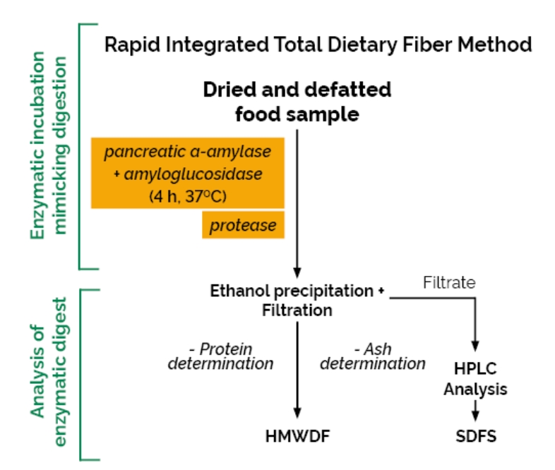
2️⃣高分子量膳食纖維 (High Molecular Weight Dietary Fiber,簡稱HMWDF),此類別又可分成
➡️➡️非水溶性膳食纖維( Insoluble Dietary Fiber,簡稱 IDF)
➡️➡️高分子量水溶性膳食纖維(水溶但78%酒精不可溶)(Soluble Dietary Fiber which precipitates in 78% ethanol,High Molecular Weight Soluble Dietary Fiber,簡稱SDFP或HMWSDF)
3️⃣低分子量水溶性膳食纖維(水溶且78%酒精也可溶)( Soluble Dietary Fiber that remains soluble in 78% ethanol,non-digestible oligosaccharides,Low Molecular Weight Soluble Dietary Fiber,簡稱SDFS或NDO或LMWSDF)
⚠️備註⚠️
➡️此套組之實驗結果,較不容易受到樣品中抗性澱粉種類(RS)和低分子量水溶性膳食纖維(LMWSDF)種類之影響
➡️低分子量水溶性膳食纖維(LMWSDF)需透過高效液相層析(HPLC)分析後,方可定量
➡️高分子量膳食纖維(HMWDF)=非水溶性膳食纖維(IDF) + 高分子量水溶性膳食纖維(SDFP)
➡️樣品中的總膳食纖維(Total dietary fiber ,簡稱TDF)=高分子量膳食纖維(HMWDF)+低分子量水溶性膳食纖維(LMWSDF)
AOAC Method 2017.16➡️此方法可分別檢測➡️高分子量膳食纖維(HMWDF)、低分子量水溶性膳食纖維(SDFS)
AOAC Method 2022.01➡️此方法可分別檢測➡️高分子量水溶性膳食纖維(SDFP)、低分子量水溶性膳食纖維(SDFS)、非水溶性膳食纖維(IDF)
.jpg)
.png)
.png)
-
樣品之酵素分解步驟(ENZYME DIGESTION OF SAMPLES)
Blanks
With each set of assays, run two blanks along with samples to measure any contribution from reagents to residue.
Samples
(a) Accurately weigh approx. 1 g sample, correct to the third decimal place, in duplicate into 250 mL Fisherbrand glass® bottles [B(b)]. Record the weight.
(b) Addition of Enzymes.— Wet the sample with 1.0 mL of 95% EtOH (or IMS) [C(a)] and add 35 mL of maleate buffer [C(g)] to each bottle. Cap the bottles. Transfer the bottles to a Grant OLS 200 shaking incubation bath (or similar) [B(g)] and secure the bottles in place with springs or polypropylene support in the shaker frame (Figure 5, page 18). Allow the solution to equilibrate to temperature for 5 min. Alternatively, use a 2mag Mixdrive 15® submersible magnetic stirrer [B(g)] with a 7 x 30 mm stirrer bar [B(p)] added to each bottle (Figure 4, page 18).
(c) Incubation with PAA/AMG solution.— Add 5 mL of PAA/AMG solution [C(d)], cap the bottles and incubate the reaction solutions at 37°C and 150 rpm in orbital motion in a shaking water bath [B(g)]; or at 170 rpm on a 2mag Mixdrive 15® submersible magnetic stirrer (to ensure complete suspension) for exactly 4 h. NOTE: If using the (NH4)2SO4 suspension of this enzyme preparation [C(d)], add 2 mL of enzyme suspension and 3 mL of sodium maleate buffer [C(g)].
(d) Adjustment of pH to approx. 8.2 (pH 7.9-8.4), Inactivation of aa-amylase and AMG.— After 4 h, remove all sample bottles from the shaking water bath and immediately add 3.0 mL of 0.75 M Tris buffer solution [C(h)] to terminate the reaction (At the same time, if only one incubation bath is available, increase the temperature of the bath to 60°C in readiness for the protease incubation step). Slightly loosen the caps of the sample bottles and immediately place the bottles in a water bath (nonshaking) at 95-100°C, and incubate for 20 min with occasional shaking (by hand). Using a thermometer, ensure that the final temperature of the bottle contents is > 90°C (checking of just one bottle is adequate).
(e) Cool.— Remove all sample bottles from the boiling water bath (use appropriate gloves) and place in the water bath set at 60°C and allow the temperature to equilibrate to approx. 60°C over 10 min.
(f) Protease treatment.— Add 0.1 mL of protease suspension (Bottle 2, page 6) [C(e)] with a positive displacement dispenser (solution is quite thick). Incubate at 60°C for 30 min.
(g) pH adjustment.— Adjust pH by adding 4.0 mL of 2 M acetic acid [C(i)] to each bottle and mix. This gives a final pH of approx 4.3.
(h) Internal standard.— Add 1.0 mL of glycerol internal standard solution (100 mg/mL; Bottle 4, page 6); [C(l)] to each incubation bottle and mix well.
(i) Proceed to step [G(a)] for determination of HMWDF (IDF + SDFP) or to step [H(a)] for determination of IDF, SDFP & SDFS.
NOTE: If available carbohydrates are to be determined, accurately transfer 0.5 mL of incubation solution to a Microfuge tube and centrifuge at 13,000 rpm for 3 min. Transfer 0.2 mL to a 13 mL (101 x 16.5 mm) polypropylene tube and add 5 mL of distilled water, cap the tube and store below -10°C awaiting analysis of available carbohydrate using the Megazyme Available Carbohydrate Assay Kit (K-AVCHO).
- DETERMINATION of HMWDF (IDF plus SDFP)
(same procedure as for AOAC Method 2009.01)
(a) Precipitation SDFP.— Preheat the sample to 60°C and add 220 mL of 95% (v/v) EtOH or IMS [C(a)] measured at room temperature and pre-heated to 60°C. Mix thoroughly and allow the precipitate to form at room temperature for 60 min (overnight precipitation is acceptable).
(b) Filtration setup.— Tare crucible containing Celite® [B(c)] to the nearest 0.1 mg. Wet and redistribute the bed of Celite® in the crucible, using 15 mL of 78% (v/v) EtOH or IMS [C(b)] from wash bottle. Apply suction to crucible to draw Celite® onto fritted glass as an even mat (Figure 6, page 19). Discard the filtrate.
(c) Filtration.— Using vacuum, filter precipitated enzyme digest [G(a)] through the crucible. Using a wash bottle with 78% (v/v) EtOH or IMS quantitatively transfer all remaining particles to crucible and wash the residue successively with two 15 mL portions of 78% (v/v) EtOH (or IMS) [C(b)]. Retain the filtrate and washings and adjust the volume to 300 mL with 78% EtOH and proceed to step [I(a)] on page 15 for determination of SDFS.
(d) Wash.— Using a vacuum, wash residue sequentially with two 15 mL portions of the following: 78% (v/v) EtOH (or IMS) [C(b)], 95% (v/v) EtOH (or IMS) [C(a)] and acetone [C(c)]. Discard these washings. Draw air through the crucible for at least 2 min to ensure all acetone is removed before drying the crucibles in an oven.
(e) Dry crucibles containing residue overnight in 105°C oven. If a forced air oven is used, loosely cover the crucibles with aluminium foil to prevent loss of dried sample.
(f) Cool crucible in desiccator for approx. 1 h. Weigh crucible containing dietary fiber residue and Celite® to nearest 0.1 mg. To obtain residue mass, subtract tare weight, i.e. weight of dried crucible and Celite®.
(g) Protein and ash determination.— The residue from one crucible is analysed for protein and the second residue of the duplicate is analysed for ash. Perform protein analysis on residue using Kjeldahl or combustion methods (Caution should be exercised when using a combustion analyser for protein in the residue. Celite® volatilised from the sample can clog the transfer lines of the unit). Use 6.25 factor for all cases to calculate mg of protein. For ash analysis, incinerate the second residue for 5 h at 525°C [B(r)]. Cool in desiccator and weigh to nearest 0.1 mg. Subtract crucible and Celite® weight to determine ash.
(h) Calculation of HMWDF (IDF + SDFP) and SDFS.— Proceed to step [ J ] (page 17).
- DETERMINATION of IDF and SDFP separately
(same procedure as for AOAC Method 2011.25)
IDF
(a) Filtration setup.— Tare crucible containing Celite® [B(c)] to nearest 0.1 mg. Wet and redistribute the bed of Celite® in the crucible, using 15 mL of 78% (v/v) EtOH (or IMS) [C(b)] from wash bottle. Apply suction to crucible to draw Celite® onto the fritted glass as an even mat (Figure 6, page 19). Discard the filtrate.
(b) Filtration.— Using vacuum, filter the enzyme digest from step [F(2)(a)] through the crucible. Using a wash bottle with 60°C deionised water rinse the incubation bottle with a minimum volume of water (approx. 10 mL) and use a rubber policeman (spatula) [B(q)] to dislodge all particles from the walls of the container. Transfer this suspension to the crucible. Wash the incubation bottle with a further 10 mL of water at 60°C and again transfer to the crucible. Collect the combined filtrate and washings and adjust the volume to 70 mL and retain this for determination of SDFP [H(f)] and SDFS [H(g)].
(c) Wash.— Using a vacuum, wash the residue successively with two 15 mL portions of the following: 78% (v/v) EtOH (or IMS) [C(b)], 95% (v/v) EtOH (or IMS) [C(a)] and acetone [C(c)]. Discard the washings. Draw air through the crucible for at least 2 min to ensure all acetone is removed (to prevent explosion hazard) before drying crucibles in oven.
(d) Dry crucibles containing residue overnight in 105°C oven.
(e) Cool crucibles and determination of IDF. Cool crucibles and determine residue mass as described in [G(e)] to [G(f)]. Determine protein and ash as described in [G(g)] and subtract from residue weight. Calculate IDF as described in step [ J ] (see NOTE, page 17).
SDFP
(f) Precipitation of SDFP.— Pre-heat the filtrate of each sample from [H(b)] (approx. 70 mL) to 60°C and add 320 mL of 95% (v/v) EtOH or IMS [C(a)] (measured at room temperature and then pre-heated to 60°C) and mix thoroughly. Allow the precipitate to form at room temperature for 60 min (overnight precipitation is acceptable).
(g) Filtration and recovery of SDFP and SDFS.— Filter the suspension and recover the residue, analyse this for protein and ash and calculate SDFP as described in steps [G(b)] to [G(g)]. Retain the filtrate and washings (approx. 420 mL) and proceed to step [I(a)] for determination of SDFS.
- DETERMINATION OF SDFS
Note: Proper deionisation is an essential part of obtaining quality chromatographic data. Refer to Figure 9 (page 20) to see patterns of glycerol and D-glucose in the presence and absence of buffer salts. To ensure that the resins being used are of adequate deionising capacity, add 0.1 mL of protease suspension (Bottle 2, page 6) to 40 mL of maleate buffer [C(g)] along with 3.0 mL of 0.75 M Tris base solution [C(h)], 4.0 mL of 2 M acetic acid [C(i)], 1 mL of glycerol internal standard (100 mg/mL; Bottle 4, page 6) [C(l)] and 1 mL of D-glucose solution (100 mg/ mL). Concentrate this solution to dryness on a rotary evaporator and re-dissolve the residue in 32 mL of deionised water. To 5 mL of this solution in a 13 mL polypropylene tube [B(s)], add 1.5 g of Amberlite® FPA53 (OH-) resin and 1.5 g of Ambersep® 200 (H+) (Figure 8, page 19), cap the tube and invert the contents regularly over 5 min. Allow the resin to settle and remove the supernatant (1.5-2.0 mL) with a syringe [B(cc)] and filter through a polyvinylidene fluoride filter, pore size 0.45 μm [B(z)]. Inject an aliquot (50 μL) of this solution onto the TSK columns (with deionising pre-column in place). A pattern similar to that shown in Figure 9c (page 20) should be obtained, i.e. no salt peaks should be evident.
(a) Filtrate recovery and concentration.— {Set aside the filtrate from one of the sample duplicates [G(c)] or [H(g)] to use if duplicate SDFS data is desired}. Transfer one quarter of the filtrate [G(c)] or [H(g)] of the second sample duplicate, {i.e. ~ 75 mL of [G(c)] or ~ 105 mL of [H(g)]} to a 500 mL evaporator flask and evaporate to dryness under vacuum at 60°C. Redissolve in 8 mL of deionised water.
(b) Deionisation of sample.— Transfer 5 mL of sample concentrate from step [I(a)] to a 13 mL polypropylene tube [B(s)] (quantitative transfer is not required as the sample contains glycerol internal standard). Add ~ 1.5 g of Amberlite® FPA53 (OH-) resin [C(p)] and ~ 1.5 g of Ambersep® 200 (H+) [C(q)] resin to the tube and invert the tube contents over 3-4 min (Figure 8, page 19).
(c) Preparation of samples for LC analyses.— Transfer the solution to a 10 mL disposable syringe [B(cc)] and filter through a 0.45 mm filter [B(z)]. Alternatively, transfer 1 mL of the solution to a microfuge centrifuge tube and centrifuge [B(ff)] at 13,000 rpm for 3 min. Use a 100 μL LC glass syringe [B(dd)] to fill the 50 μL injection loop on the LC [B(t)]. A single analysis of the sample is adequate. Columns: Two TOSOH TSK gel permeation columns [B(w)] with deionising pre-column [B(v)]. Solvent: microfiltered [B(bb)], distilled water. Flow rate: 0.5 mL/min; 60 min per run. Temperature: 80°C, Figure 7, page 19).
(d) Determine the response factor for D-glucose.— Because D-glucose provides an LC refractive index (RI) response equivalent to the response factor for the non-digestible oligosaccharides that make up SDFS, D-glucose is used to calibrated the LC and the response factor is used for determining the mass of SDFS. Use a 100 μL LC syringe to fill a 50 μL injection loop with the D-glucose/glycerol internal standard solution (Bottle 5, page 6). Inject in duplicate. Calculate the response factor according to [J(b)(1)].
(e) Calibrate the area of chromatogram to be measured for SDFS.— Use a 100 μL LC syringe [B(dd)], to fill the 50 μL injection loop with retention time standard Bottle 3 (page 6) [C(m)]. Inject in duplicate. Determine demarcation point between DP 2 and DP 3 oligosaccharides (disaccharide maltose versus higher oligosaccharides) (Figure 3, page 4).
(f) Determine peak area of SDFS (PASDFS) and internal standard (PAIS) in chromatograms of sample extracts.— Inject sample extracts [I(c)] on LC. Record area of all peaks of DP greater than the DP2/DP3 demarcation point as PASDFS. Record the peak area of internal standard as PAIS. (g) Proceed to step [ J(b)(2)].
商品特色
商品規格
- 商品規格(100個檢測反應)
- 100個檢測反應套組(100 determinations assay kit)
Bottle 1: (*2)
Mixture of purified PAA (40 KU/g) and AMG (17 KU/g) pancreatic a-amylase; 5.2 g.
Stable for > 5 years stored dry below -10°C.
NOTE: For some individuals, these powdered enzymes are highly allergenic. Thus, they should be weighed and handled ONLY in a fume cupboard; see [C(d)].
Bottle 2:
Purified protease (E-BSPAMS) (10.5 mL, 350 tyrosine U/mL in 3.2 M ammonium sulphate).
Stable for > 3 years at 4°C.
Bottle 3:
LC Retention Time Standard [maltodextrins plus maltose (4:1 ratio)], approx. 5 g.
Stable for > 3 years; store sealed at room temperature.
Bottle 4: (*2)
Glycerol standard solution (100 mg/mL, 55 mL).
Stable for > 4 years; store sealed at 4°C.
Bottle 5:
D-Glucose/glycerol (Glu/Gly) standard solution (10 mg/mL of each in 0.02% w/v sodium azide).
Stable for > 4 years; store sealed at 4°C.